Retinal Detachment
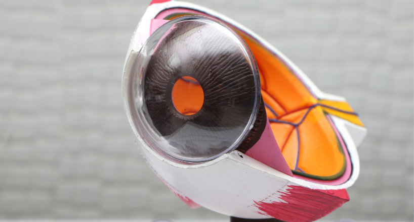
A retinal detachment is a major condition in which the retina, the light-sensitive layer of tissue at the back of the eye, becomes separated from the underlying layers of the eye. The retina is responsible for converting light into electrical signals that the brain translates as vision. When the retina ends up being removed, it can no longer operate effectively, resulting in vision loss.
There are 3 main types of retinal detachment:
- Rhegmatogenous retinal detachment: This is the most common kind of detachment and it occurs when a tear or hole in the retina enables fluid to permeate underneath and separate the retina from the underlying layers of the eye.
- Tractional retinal detachment: This type of detachment occurs when scar tissue on the surface of the retina triggers it to retreat from the underlying layers of the eye.
- Exudative retinal detachment: This type of detachment happens when fluid leakages into the space in between the retina and the underlying layers of the eye as an outcome of a hidden condition such as diabetes or age-related macular degeneration.
Signs and Symptoms
Symptoms of retinal detachment consist of a sudden onset of floaters (tiny spots or cobweb-like shapes that wander throughout the field of vision), flashing lights, and a dark or empty area in the field of vision. Retinal detachment is a medical emergency situation and needs instant treatment to prevent irreversible vision loss. Treatment alternatives include laser surgical treatment, cryotherapy, or surgical repair.
Causes and Risk Factors
Retinal detachment can be brought on by an injury to the eye or face, as a result of diabetic retinopathy or extremely high nearsightedness (in which the retina is thinner than in normal eyes). It can also arise from modifications in the vitreous of the eye due to aging, eye or other systemic diseases, or following eye surgery.
Elements that put you in danger increased to consist of:
- Age- a retinal detachment is more typical in grownups 50 and over
- Diabetes or Sickle Cell
- Severe nearsightedness
- Eye surgical treatment (such as cataract elimination)
- Eye or face injury
- Family history
- Eye disease or inflammation
Treatment for Retinal Detachment

Treatment for retinal detachment usually involves surgical repair of the retina.
The kind of surgery utilized will depend upon the type of retinal detachment and the underlying cause.
The main surgical choices for retinal detachment include:
- Cryopexy: This is a treatment in which a freezing probe is utilized to seal a retinal tear or hole.
- Laser photocoagulation: This is a treatment in which a laser is utilized to develop small burns around the retinal tear or hole, which helps to seal it and prevent further fluid from seeping underneath the retina.
- Pneumatic retinopexy: This is a treatment in which a gas bubble is injected into the eye to press the retina back into place.
- Vitrectomy: This is a procedure in which the vitreous gel is gotten rid of from the eye, along with any scar tissue that is pulling on the retina. A gas or silicone bubble is then utilized to hold the retina in place while it heals.
Sometimes, a mix of these treatments might be used.
It is likewise essential to note that after the surgical treatment, the patient will need to preserve a particular position for a few days, this is referred to as posturing. The kind of posturing will depend upon the type of surgery carried out, the surgeon will supply guidelines to the patient.
In some cases, retinal detachment may not be repairable and the vision loss might be long-term. Early detection and treatment are key to minimizing the danger of vision loss.