To understand how the human eye works, initially picture a photographic camera– considering that cams were established very much with the human eye in mind.
How do we see what we see?
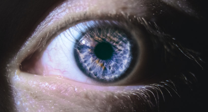
Light reflects off of items and gets in the eyeball through a transparent layer of tissue at the front of the eye called the cornea. The cornea accepts extensively divergent light rays and flexes them through the student– the dark opening in the center of the colored part of the eye.
The student appears to broaden or contract instantly based on the strength of the light going into the eye. In fact, this action is controlled by the iris– a ring of muscles within the colored portion of the eye that adjusts the pupil opening based on the strength of light. (So when a student appears to broaden or agree, it is in fact the iris doing its task.)
The adjusted light travels through the lens of the eye. Located behind the pupil, the lens automatically changes the path of the light and brings it into sharp focus onto the getting location at the back of the eye– the retina.
A fantastic membrane filled with photoreceptors (a.k.a. the “rods and cones”), the retina transforms the light rays into electrical impulses. These then take a trip through the optic nerve at the back of the eye to the brain, where an image is finally perceived.
A fragile system, based on flaws.
It’s easy to see that a slight change in any element of how the human eye works — the shape of the eyeball, the cornea’s health, lens shape and curvature, and retina issues– can trigger the eye to produce fuzzy or blurred vision. That is why many people need vision correction. Spectacles and contact lenses assist the light focus images correctly on the retina and enable individuals to see clearly.
As result, a lens is put in front of the eye to make up for any shortages in the complex vision process.
The main parts of the human eye include:
- Cornea: transparent tissue covering the front of the eye that lets light travel through
- Iris: a ring of muscles in the colored part of the eye that manages the size of the pupil
- Pupil: an opening in the center of the iris that alters size to control just how much light is getting in the eye.
- Sclera: the white part of the eye that is composed of fibrous tissue that secures the inner functions of the eye
- Lens: located directly behind the pupil, it focuses light rays onto the retina
- Retina: membrane at the back of the eye that alters light into nerve signals
- Rods and cones: unique cells utilized by the retina to procedure light
- Fovea: a tiny area in the center of the retina that contains just cone cells. It permits us to see things sharply.
- Optic Nerve: a bundle of nerve fibers that carries messages from the eyes to the brain
- Macula: a small and highly delicate part of the retina responsible for main vision, which enables an individual to see shapes, colors, and details clearly and sharply.